
 Driven by curiosity. Driving change
Our equipment
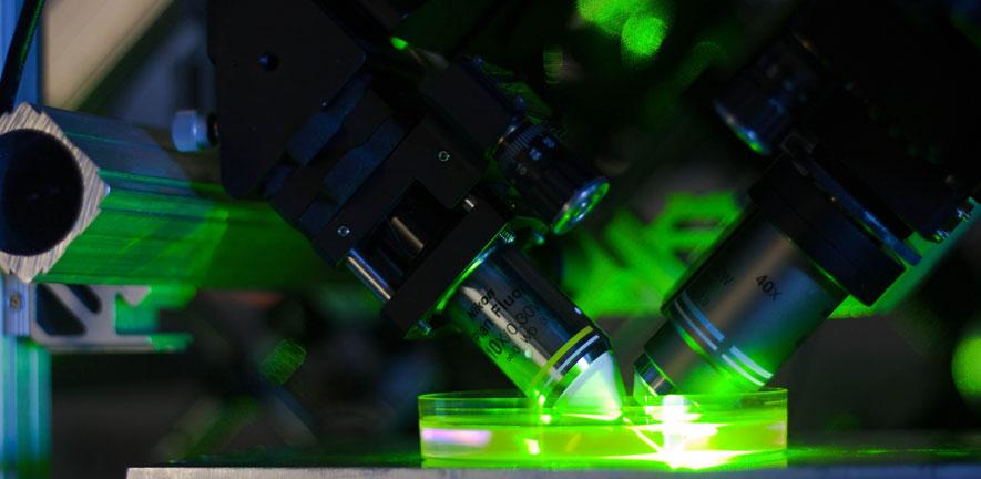
Our facilities include optical super-resolution microscopes using variants of single molecule localisation microscopy such as dSTORM and DNA-PAINT, structured illumination microscopy (SIM) systems and stimulated emission depletion (STED) microscope systems. We have a three-channel oblique plane microscopy (OPM) system for high-speed and high-resolution volumetric imaging. Furthermore we have systems for whole organism imaging methods such as selective plane illumination microscopy. These efforts were funded over the past 10 years by grants from the Wellcome Trust, MRC, EPSRC, BBSRC and the Alzheimer Research Trust UK.
We have state-of-the-art laser sources and detector systems including several supercontinuum laser sources, Ti:Sa lasers, EMCCD and scientific-grade CMOS cameras, microscopic imaging stations etc. We also have state of the art molecular biology, cell culture and chemical preparation laboratories.
Key equipment is listed below:
Equipment
Structured Illumination Microscope (SIM)
In our lab we have developed a SIM microscope capturing super-resolution images with, a resolution twice as high as for confocal imaging, in fractions of a second. The system can operate in TIRF or widefield SIM modes with simultaneous multicolour excitation – allowing the system to be flexibly adapted to the imaging problem.
Widefield time-gated fluorescence lifetime imaging microscope (TG-FLIM)
We have developed a widefield time-gated FLIM microscope capable of taking FLIM images rapidly, as fast as 2Hz. The system is equipped with an automated stage and our home-built LabView script allows automation and unsupervised, rapid screening of samples
Oblique Plane Microscopy (OPM)
We have developed a high-NA oblique plane microscopy system that uses the glass-tipped AMS-AGY v1 objective lens (a.k.a. Snouty lens) as the tertiary objective lens, a three-channel emission splitter (Cairn Optosplit 3) and a Photometrics Kinetix sCMOS camera.
Direct Stochastic Optical Reconstruction Microscopy (dSTORM)
We have a custom built dSTORM microscope, capable of simultaneous two-colour super-resolution imaging and we achieve an image resolution of 15 nm routinely.
DNA Points Accumulation for Imaging in Nanoscale Topography (DNA-PAINT)
We are using a custom built setup for DNA-PAINT, capable of multiplexed super-resolution imaging with a resolution of about 40 nm. We are currently working on several approaches to enhance acquisition speed and multi-channel imaging.
Stimulated Emission Depletion Microscope (STED)
We have developed a state of the art STED microscope for fast imaging of samples labelled with red dyes such as the ATTO or Aberrior dyes. The STED system uses a Ti:Sa laser beam which is spatially shaped by an SLM to increase the effective resolution of a standard confocal microscope from 250 nm to 30-90 nm. This technique works at reasonable imaging speed and depths and works well to image molecules deep within cells.
Selective Plane Illumination Microscope (SPIM)
We have developed a state-of-the-art SPIM microscope for high speed single-cell imaging. The system is capable of recording up to 60 fluorescence sections per second and is mostly used for live single-cell and expanded sample imaging.
Confocal imaging station for multi-parameter imaging (TCSPC)
We have developed a confocal microscopy based platform integrated with a time correlated single photon counting (TCSPC) module, for fluorescence lifetime imaging measurements. The system employs either a pulsed femtosecond laser (for two-photon (2P) measurements) or a supercontinuum laser, for excitation at repetition rates ranging 2-80 MHz. Hence, it is unique in terms of its flexibility of use and sensitivity, especially for biological imaging.
Cold Scope
The group has recently developed a method for high-fidelity imaging of live biological samples at temperatures of around, or below, 0 °C. It relies on hardware additions to traditional microscopy, namely as a cooling collar. It can be straightforwardly implemented on different microscopy modalities. We have been regularly using it on our SIM and SPT systems.
Atomic Force Microscopy
The group also has a state-of-the-art atomic force microscope (AFM) for life science and materials science research. AFM exploits the use of sharp probes for the creation of ultra-high resolution maps of surface topography alongside physical properties maps of the sample, such as elasticity, adhesion etc. The instrument in our lab, (a Bruker Resolve AFM) can be easily integrated with most inverted microscope frames. This allows for the development of correlative microscopy platforms, such as AFM-STED, AFM-FLIM and AFM-SIM.
Molecular and cell biological facilities
We have state of the art molecular biology, class II cell culture and nematode facilities. The facilities include equipment to perform protein purification, molecular cloning, bio-chemical and -physical assays, and mammalian cell transfection.